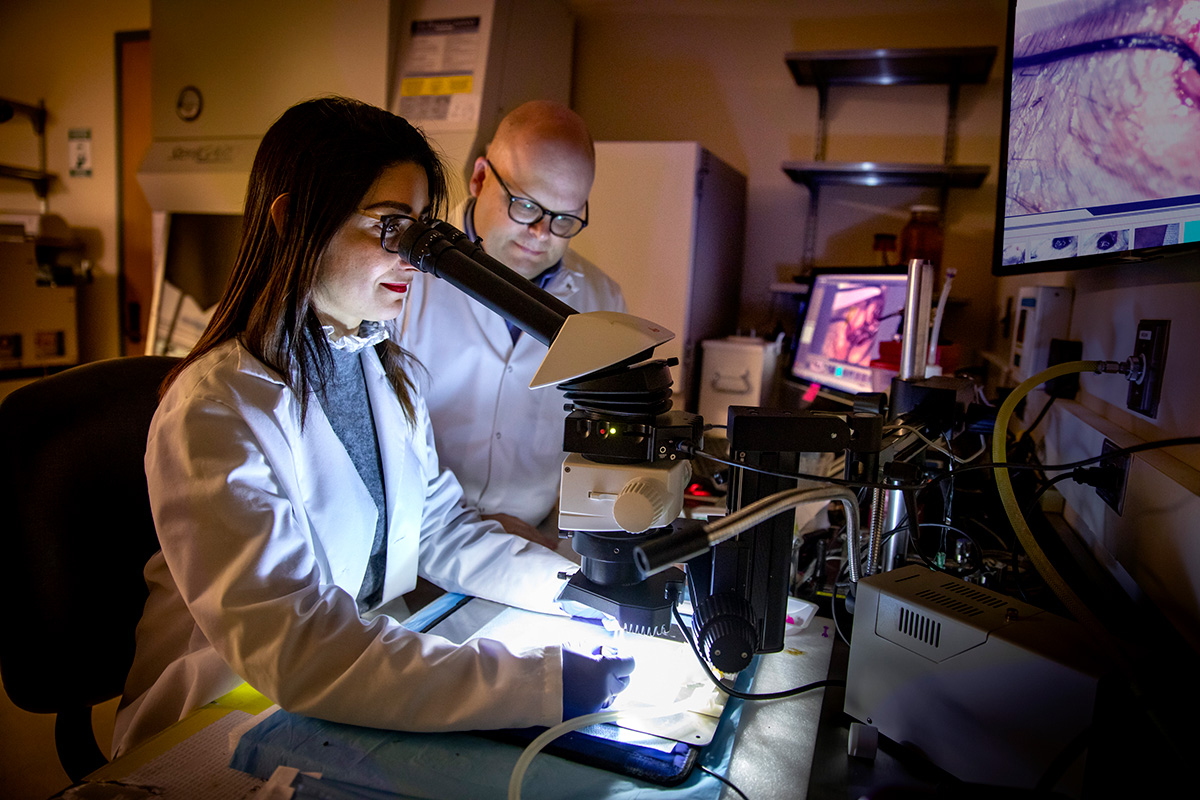
 Since Galileo’s time, imaging has served as the visionary guide of scientific discovery. Fast forward centuries later, the National Academy of Engineering hailed imaging as one of the most remarkable engineering feats of the 20th century. Now, imagine the incredible potential that lies within combining various imaging techniques and technologies to map the intricate biological processes happening within a single cell or even entire organs. This breakthrough has the power to completely revolutionize the way we diagnose and treat pathophysiological disorders, ultimately reducing the staggering social and economic burdens associated with disease management. At EMIL, our research endeavors are dedicated to creating these groundbreaking integrated imaging approaches, paving the way for personalized disease prevention programs through advanced diagnosis, precise risk assessment, and targeted cell therapies.
Theme One: Noninvasive Monitoring of Angiogenesis
Picture a world where we can unlock the secrets of angiogenesis, the captivating process of creating new blood vessels. This phenomenon holds immense significance in the realm of common ailments like myocardial ischemia, coronary artery disease (CAD), peripheral arterial disease (PAD), and even cancer. Leading academic institutions worldwide are fervently pursuing therapeutic strategies aimed at stimulating angiogenesis through gene therapy and stem cells. Yet, despite promising outcomes in animal studies, clinical trials attempting to stimulate angiogenesis in CAD and PAD have left us scratching our heads, as they showed no clear advantages over placebos.
Here’s where things get interesting: the realm of noninvasive imaging approaches to track these vital biological processes remains largely uncharted. That’s where our groundbreaking research comes in. We are dedicated to developing cutting-edge multimodal imaging strategies that harness the power of nuclear imaging (PET, SPECT, X-ray CT) and optical imaging (fluorescence and luminescence) to delve deep into the cardiovascular mysteries of pathologies like peripheral arterial disease (PAD). Our goal? Noninvasively monitor the mesmerizing dance of angiogenesis and uncover its secrets.
But we don’t stop there. We’re also investigating the molecular targets crucial to vessel formation and maturation, with the potential to serve as targeted tracers for monitoring therapeutic interventions. In fact, we’ve already shown promising results in our previous studies, where we demonstrated the feasibility of gene transfer using intramyocardial insulin-like growth factor (IGF-1) after a heart attack, resulting in sustained angiogenesis and improved heart function. But it doesn’t end with gene therapy. We’re also exploring the captivating world of cell-based approaches, including transplanting stem cells, exosomes, or bioengineered scaffolds populated with cells. These innovative techniques hold immense potential.
Right now, our focus is on the development, radiosynthesis, and characterization of cRGD-peptide constructs. These incredible tools allow us to non-invasively track the captivating angiogenic processes in preclinical models of PAD and the post-infarct myocardium during the onset of diabetes. We’re taking imaging to a whole new level, unraveling the intricate story of angiogenesis and paving the way for transformative advancements in the field of cardiovascular health.
Theme Two: Imaging the Receptor for Advanced Glycation End-products (RAGE)
 The receptor for advanced glycation end-products (RAGE) is a receptor with an immunoglobulin-like structure located in the cell membrane. It has multiple isoforms that enable it to bind to various types of endogenous and exogenous ligands. When these ligands bind to RAGE, it triggers a series of complex pathways that lead to the production of free radicals like reactive nitric oxide and oxygen species, cell proliferation, and immunoinflammatory processes.
The role of RAGE in the development of disorders such as diabetes, inflammation, tumor progression, and endothelial dysfunction is influenced by the accumulation of advanced glycation end-products (AGEs) in pathological conditions, resulting in increased expression of RAGE. Given the involvement of RAGE and its ligands in numerous diseases and pathologies, targeting RAGE through therapies that modulate its expression or activation, as well as the production or administration of AGEs, is of great interest. However, despite the known significance of the RAGE/AGE axis in multiple diseases, there is an urgent need to develop non-invasive molecular imaging methods that can accurately measure RAGE levels in living organisms. Such approaches would greatly contribute to validating RAGE and its ligands as biomarkers and potential targets for therapeutic interventions.
At EMIL, we specialize in the development of nanoparticle-based and peptide-based constructs. These constructs can be labeled with radioisotopes or fluorescent markers, allowing for non-invasive tracking of the RAGE/AGE axis using PET-CT or optical imaging techniques.
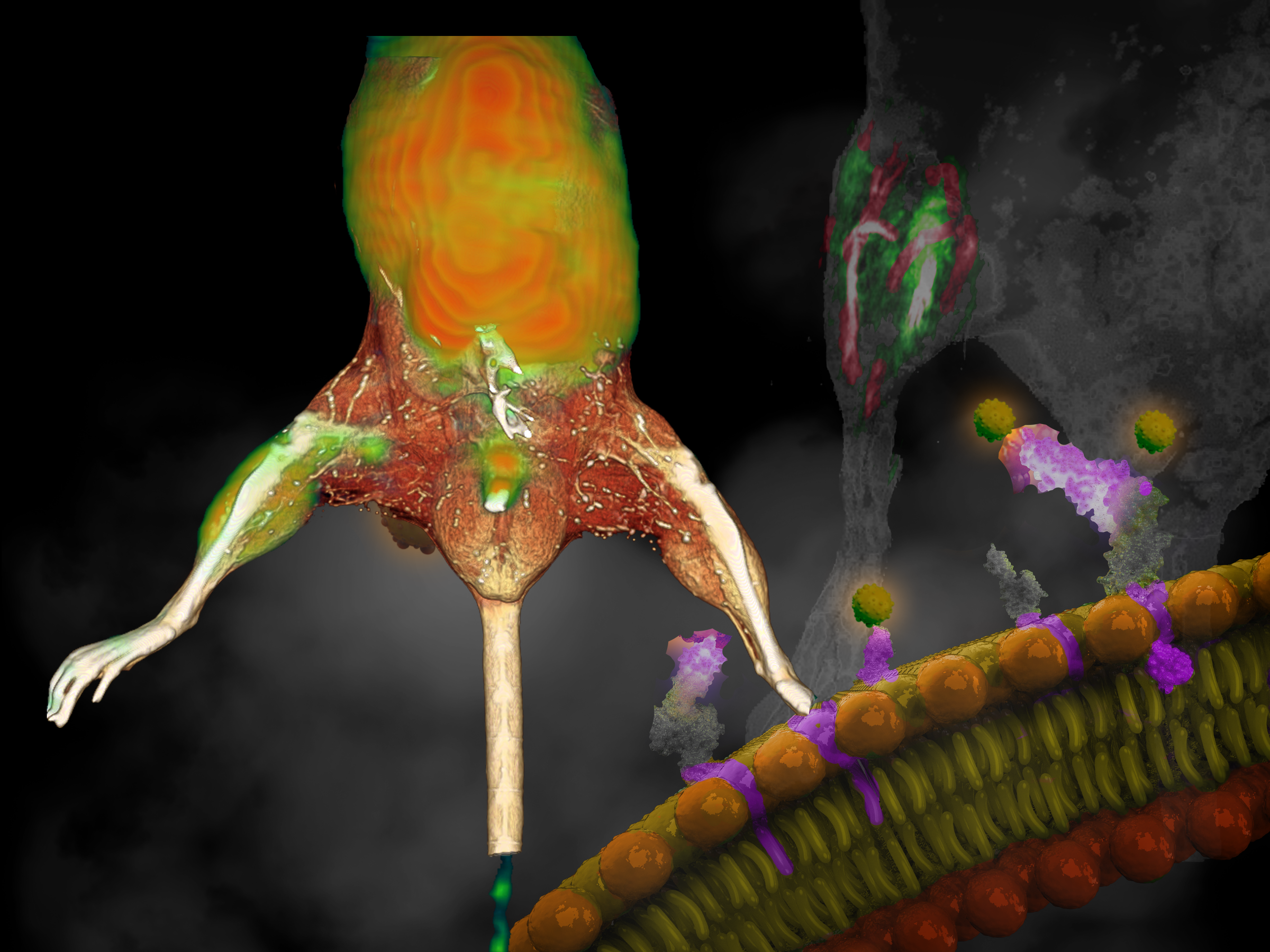
Theme Three: Development of Multimodal Multifunctional Targeted Tracers
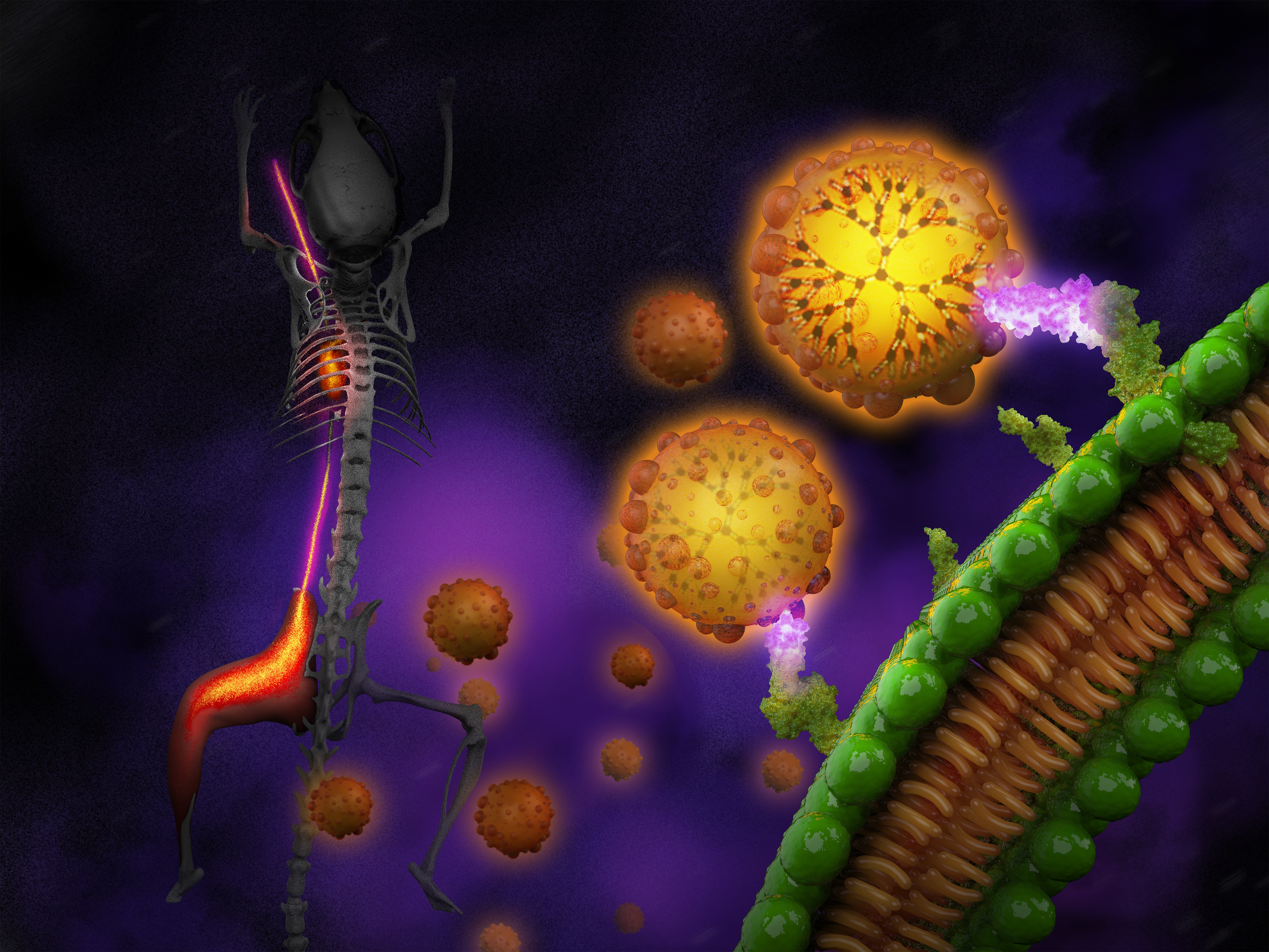
With the introduction of molecular probes, the use of imaging techniques now allows us to analyze the time-based and spatial distributions of biological processes. To advance the field of hybridized systems, it is necessary to have innovative imaging agents that are capable of multiple modes and functions, enabling the study of biology in vivo not only at the level of individual components but also in terms of system processes and mechanisms. These agents can be further utilized for computer simulations. Furthermore, by combining the imaging of biological processes with personalized therapies, we can eventually create novel modular and multifunctional nanoparticle structures that facilitate targeted therapy delivery alongside multimodal imaging. Our previous research focused on developing dendrimer-based probes for imaging the blood pool, which led us to design modular elements that can be manipulated to create an optimized multifunctional platform. These modular elements encompass liposomes for encapsulation, biodegradable solid particles for sustained drug delivery, and dendrimer nanoparticles or quantum dots for multimodal labeling. If successful, this multifunctional approach will bring about a significant shift in our understanding and management of patients, introducing a completely new diagnostic and therapeutic approach.